Best anatomy yokochi
If you looking for anatomy yokochi then you are right place. We are searching for the best anatomy yokochi on the market and analyze these products to provide you the best choice.
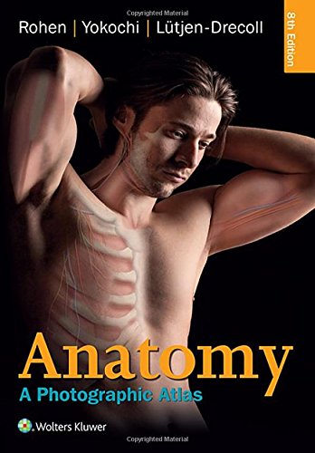


1. Anatomy: A Photographic Atlas (Color Atlas of Anatomy a Photographic Study of the Human Body)
Description
Prepare for the dissection lab and operating room with Anatomy: A Photographic Atlas, 8e. Featuring outstanding full-color photographs of actual cadaver dissections with accompanying schematic drawings and diagnostic images, this proven text depicts anatomic structures more realistically than illustrations in traditional atlases. Chapters are organized by region in the order of a typical dissection with each chapter presenting topographical anatomical structures in a systemic manner.
- Authentic photographic reproduction of colors, structures, and spatial dimensions as seen in the dissection lab and on the operating table help you develop an understanding of the anatomy of the human body.
- Functional connections between single organs, the surrounding tissue, and organ systems are clarified to prepare you for the dissection lab and practical exams.
- Clinical cases and over 1,200 images enhance your understanding.
- Dissections illustrate the topographical anatomy in layers "from the outside in" to better prepare you for the lab and operating room.
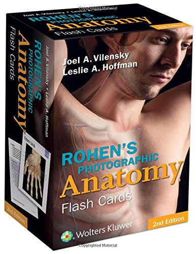
2. Rohen's Photographic Anatomy Flash Cards
Feature
MPN: 9781451194500Authentic Anatomical Chart Company product!
Measures 7 x 5 inches
Great for studies and patient consultation
Printed in the United States
Description
- Now includes review questions and additional clinical anatomy.
- 220 full-color photographs present exquisitely dissected cadavers.
- Ideal study aid to review recently dissected regions and to prepare for practical exams.
- Includes a key ring and hole-punched corners for easy organization and on-the-go portability.
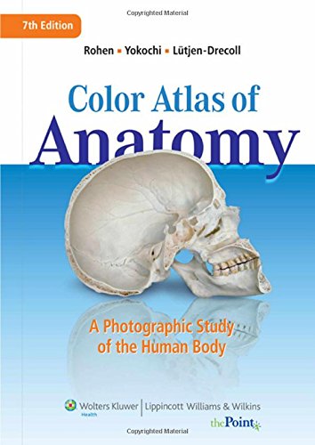
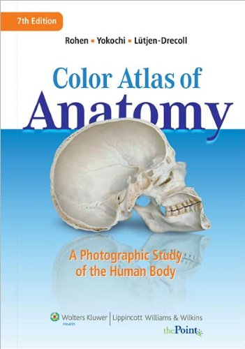
3. Color Atlas of Anatomy: A Photographic Study of the Human Body
Feature
Used Book in Good ConditionDescription
This Color Atlas of Anatomy features full-color photographs of actual cadaver dissections, with accompanying schematic drawings and diagnostic images. The photographs depict anatomic structures with a realism unmatched by illustrations in traditional atlases and show students specimens as they will appear in the dissection lab.
Chapters are organized by region in order of standard dissection, with structures presented both in a systemic manner, from deep to surface, and in a regional manner.
This edition has additional clinical imaging, including MRIs, CTs, and endoscopic techniques. New graphics include clinically relevant nerve and vessel varieties and antagonistic muscle functions. Many older images have been replaced with new, high-resolution images. Black-and-white dissection photographs have been replaced with color photography.
A companion website will include an Image Bank, interactive software (similar to an Interactive Atlas), and full text online.
4. Color Atlas of Anatomy: A Photographic Study of the Human Body (Color Atlas of Anatomy (Rohen))
Feature
Crisp color pictures (cartoons, real cadavor, and x-ray imaging)Description
This atlas features outstanding full-color photographs of actual cadaver dissections, with accompanying schematic drawings and diagnostic images. The photographs depict anatomic structures more realistically than illustrations in traditional atlases and show students exactly what they will see in the dissection lab.
Chapters are organized by region in order of a typical dissection. Each chapter presents structures both in a systemic manner from deep to surface, and in a regional manner.
This edition has sixteen additional pages of clinical imagesincluding CT and MRIthat students can compare with cross-sectional anatomic photographs. Many pictures have been electronically enhanced or rescanned for better contrasts.
5. Color Atlas of Anatomy: A Photographic Study of the Human Body
Feature
Used Book in Good ConditionDescription
The on-going core of this atlas is its standard of realistic illustrations that portray anatomical relationships. Photographs of actual cadaver dissections along with numerous schematic drawings aid the student in anatomic orientation. Chapters are organized by region, in order of a typical dissection. Each chapter contains two sections: a description and illustration of organs, and a depiction of those organs within the regional anatomy. New to this edition is an increase of MRI pictures, approximately 30 schematic drawings made even more precise, and an updated text where appropriate.
A Brandon-Hill recommended title.
6. Photographic Anatomy of the Human Body
Description
Softcover atlas consisting of high-quality photographic color plates, for allied health students. DNLM: 1. Anatomy - atlases.7. Color Atlas of Anatomy (text only) 7th (Seventh) edition by J. W. Rohen,E. Ltjen-Drecoll,C. Yokochi
Description
Color Atlas of Anatomy: A Photographic Study of the Human Body (Point (Lippincott Williams & Wilkins)) [Hardcover]Johannes W. Rohen (Author) , Elke Ltjen-Drecoll (Author), Chichiro Yokochi (Author)8. Rohen's Photographic Anatomy Flash Cards
Description
This set of 220 flash cards is based on the images in Color Atlas of Anatomy: A Photographic Study of the Human Body, Sixth Edition. This is the only gross anatomy flash card set that includes full-color photographs of actual cadaver dissections. The photographs realistically depict anatomic structures as seen on the cadaver, allowing students to prepare for lab dissections and study for practical laboratory exams.
On the front of each card is an image with key structures labeled. On the back of each card are hints to help identify the structure and relevant clinical pearls.
9. Color Atlas of Anatomy: A Photographic Study of the Human Body by Johannes W. Rohen (1998-01-15)
Description
Color Atlas of Anatomy: A Photographic Study of the Human Body by Johannes W. Rohen (1998-01-15)10. Color Atlas of Anatomy: A Photographic Study of the Human Body 7th (seventh) revised internat Edition by Johannes W. Rohen, Elke Lutjen-Drecoll, Chichiro Yokochi published by Lippincott Williams and Wilkins (2010)


Recent Comments